
Mazzulo's Market: Caterers Aurora, OH - aurora oh
In the intricate world of medical imaging, ultrasound machines stand out as non-invasive, versatile tools for diagnostic and therapeutic purposes. Understanding the ultrasound machine parts and functions, from the crucial ultrasound probe parts to the sophisticated ultrasound machine components like the CPU and software, is essential for healthcare professionals. Each part, whether it's the gel dispenser that ensures optimal sound transmission or the foot pedals that provide hands-free operation during critical procedures, plays a vital role in the functionality and efficiency of the ultrasound equipment.
The display screen is where the magic happens—where ultrasound images come to life. These screens are engineered to deliver high-resolution, real-time images that allow medical professionals to visualize the patient's internal structures. Modern ultrasound scanners frequently have color displays with many viewing modes, including 2D, 3D, and even 4D (real-time 3D), which provide detailed and dynamic pictures of the body's structure.
XityMedical people always uphold the craftsman spirit, and are renowned for quick response, professional services and reasonable prices in the medical industry.
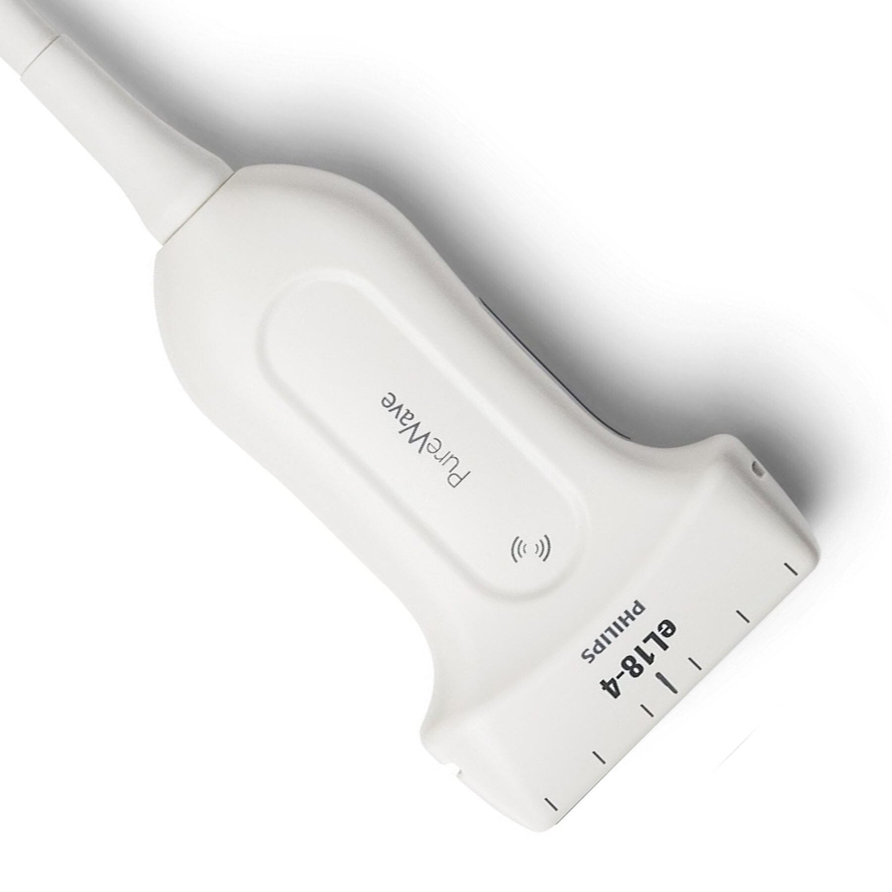
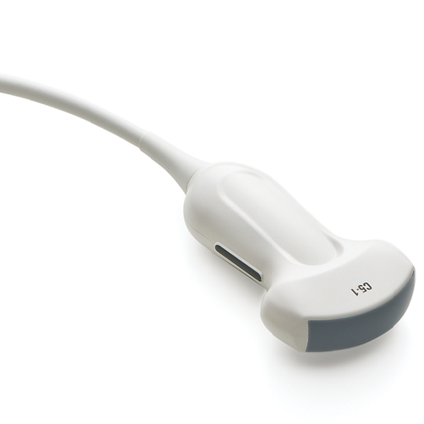
The ultrasound transducer is not just the heart, but the core of the ultrasound machine, playing a pivotal role in parts of ultrasound imaging. This handheld device is adept at emitting high-frequency sound waves and capturing the echoes that reflect off bodily structures. The transducer houses sophisticated piezoelectric crystals, which are the essence of ultrasound technology. Upon receiving electrical current, these crystals oscillate, sending sound waves deep into the body. The reflected echoes are then captured by the same transducer, which converts them into electrical signals. These signals are processed by the machine's CPU to produce the live images we see on the display screen.
For those in the medical field, recognizing the importance of each component, including the ultrasound machine buttons and functions, can lead to more accurate diagnoses and better patient care. As a leading provider of medical imaging solutions, Xity offers an extensive array of ultrasound equipment parts and accessories designed to meet the diverse needs of healthcare practitioners. Our selection ranges from the basic components of ultrasound machines to advanced parts of the ultrasound probes, ensuring that your sonography machine operates at its best.
Foot pedals provide operators with a hands-free way to control certain functions of the ultrasound machine. They are particularly useful during dynamic procedures when the operator needs to freeze or capture specific moments during an examination without interrupting the scan. Foot pedals improve accuracy and control, ensuring that critical photos are caught when needed.
Let us pre-assemble and inspect the quality for you. We follow the IPC standards and nothing leaves the door unless the quality meets Class 2 or higher. Not only can you cut down on the extra cost associated with subcontracting the secondary operation, it’s one less thing you have to worry about.
If you're looking to enhance your ultrasound machine's capabilities or require detailed information on parts of an ultrasound machine like the 3D Motor Controllers, Channel Board, Power Regulator, and more, Xity is here to assist. Reach out to us for expert advice and premium ultrasound machine parts tailored to your specific medical imaging requirements.
Ultrasound gel is a conductive medium that helps transmit sound waves between the transducer and the patient's skin. The gel dispenser is a convenient feature built into many ultrasound machines, ensuring that the gel is readily available during the examination. This gel enhances image quality by minimizing air gaps between the transducer and the patient's body, resulting in better acoustic coupling and clearer imaging of inner structures.
Behind the scenes, the ultrasound machine is powered by a robust central processing unit (CPU) and specialized software. The CPU is responsible for processing the incoming data from the transducer and converting it into visual images displayed on the screen. The software, on the other hand, plays a critical role in image enhancement and manipulation. In the case of 3D/4D images, it may apply filters, modify contrast, and even generate three-dimensional reconstructions.
Transducers vary in shape and size to cater to different diagnostic requirements. For instance, parts of an ultrasound probe designed for superficial structures, like linear transducers, differ from those meant for deeper examinations, such as curved transducers used in abdominal scans. Understanding the ultrasound probe parts and their specific functions is crucial for medical practitioners to select the appropriate transducer for each diagnostic scenario.
The control panel is where the operator, usually a sonographer or a radiologist, adjusts various settings of the ultrasound machine to optimize image quality. It has controls for factors including frequency, depth, gain, and focus through buttons, knobs, and a keyboard. These settings can be adjusted to obtain the best possible images for different types of examinations.
2. The specification table above are summarised information, so please confirm further details on the respective detail specification.
The clarity and precision of the pictures produced by the machine are directly affected by the quality of the display screen, which is critical for correct diagnosis.
The probe connector is the physical interface that connects the transducer to the ultrasound machine. It ensures that data from the transducer, including the emitted ultrasound waves and received echoes, is accurately transmitted to the CPU for processing. The ability to switch between different types of probes is critical because it allows healthcare providers to adjust the equipment to varied clinical settings and patient demands.
Noritake Itron offers 5 colors (gray, blue, green, aqua and rose) in 11 popular VFD module display sizes. Additional colors and sizes are available upon request
There is currently a code library that provides access to the basic functionality of the GU7000 series modules using 8-bit Atmel AVR or Linux. This library is intended for use with the following modules:
≫ Request a Demo or Make a Purchase ≫ View the entire GU-7000 Product Line-up General Description ≫ GU-7000 Application Note Code Library Available ≫ Noritake_VFD_GU7000 Demo Files Available ≫ Noritake_VFD_GU7000_LargeTextDemo ≫ Noritake_VFD_GU7000_ImageDemo Support Tools Available ≫ Bitmap Image Tool ≫ Text Encoding Converter ≫ Bitmap Image Loader
Ultrasound machines, also known as sonography machines, are essential tools in the field of medicine and have revolutionized the way healthcare professionals diagnose and monitor various medical conditions. These machines use high-frequency sound waves to create real-time images of the inside of the body. In this post, we will look at the main components of an ultrasound machine and how they work.


 Neil
Neil 
 Neil
Neil