
Lithium cells & batteries - 3.0 v lithium battery
De moord op Jacques d'A.: Directed by Hans de Korte. With John Jones, Angelique de Bruijne, Anneke Beukman, Jules Royaards.
The Bay 1 area contains the necessary subject care environment including waiting and changing rooms; support areas including a business office; a data viewing area; a physician’s office; and computer and magnet rooms.
The 11.7 Tesla laboratory includes a 54 mm i.d. vertical bore, actively screened magnet interfaced to a JEOL 500 MHz NMR console with variable temperature capability. This instrument has 3 RF channels, each with complete waveform shaping capability. Channel 1 is highband, with 1H/19F observe, spinlock and decouple capability (100 W, 455-535 MHz). Channel 2 has broadband observe, spinlock and decouple capability (10-210 MHz, 300 W). Channel 3 is highband, with 1H/19F observe, spinlock and decouple capability (100 W, 455-535 MHz). The probe consists of a 5mm ROYAL HFX, triple-channel NMR probe (1H,19F, and X) with 3G/A*cm Z gradient, and BB range from 31P to 109Ag. The probe is auto tune/matched and has VT capabilities for operation between -170 to +250°C.
This system has EPI, second order shimming, CINE, MR angiography, diffusion, perfusion, and spectroscopy capabilities for both neuro and body applications. It uses the same gradients as the 1.5 T Avanto (Bay 2; 45 mT/m strength, 200T/m/s slew rate).
The 14.1 Tesla (600MHz) laboratory consists of a 8.9-cm wide bore, actively screened, vertical bore Magnex magnet interfaced to a Bruker Avance III HD spectrometer console. Capabilities include dual RF channels and deuterium lock; 5-mm and 10-mm direct and indirect observation high-resolution (0.7 ppb) multinuclear multidimensional liquid state spectroscopy; high-resolution (1.6 ppb) 1H and 13C MAS spectroscopy (including gradient spectroscopy); high-power multinuclear cross polarization/magic angle spinning (CP/MAS) spectroscopy; an automated MAS sample changer; multinuclear (31P/1H, 19F/1H, and 13C/1H) micro-imaging and in vivo spectroscopy; actively screened gradients with up to 150 G/cm; and variable controlled temperature from –100 to +150 °C with stability approaching 0.1 °C.
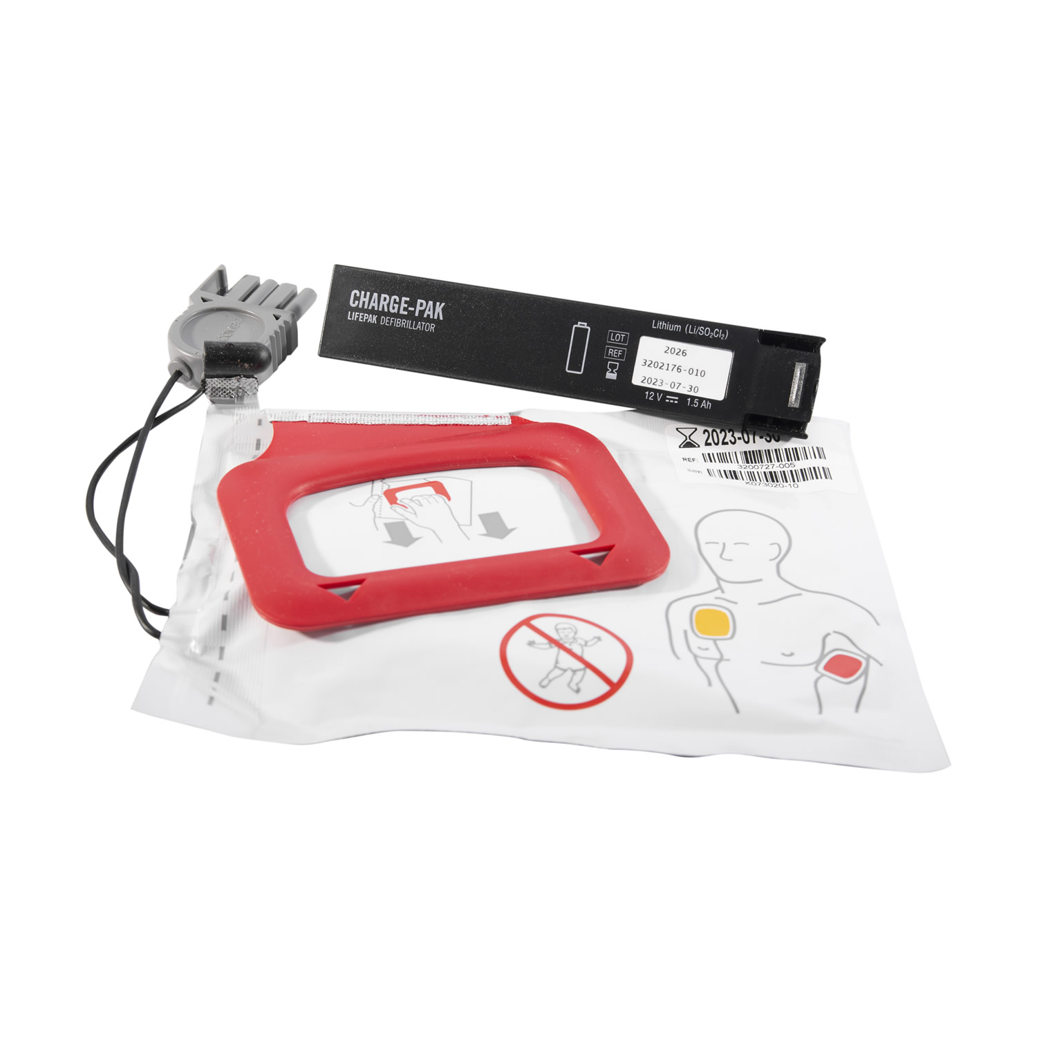
With a stand-by life of 2 years and the same replace-by date, the Physio-Control Lifepak CR Plus CHARGE-PAK and electrode kit provides a complete pack to easily replace components that have expired or have been used. Available to purchase with 1 or 2 pairs of QUICK-PAK electrodes, the replacement kit is designed for use in conjunction with the Physio-Control Lifepak CR Plus Fully Auto and Semi-Auto Defibrillators.
This is a 32-channel Siemens Tim Trio 3T whole-body MRI scanner with an insertable 36-cm (gradient coil ID) head-only gradient. The whole-body gradient system uses the same gradients as the 1.5T Avanto (45 mT/m strength, 200T/m/s slew rate). It has 32 independent RF receive channels for phased array coils, including a Siemens 32-channel head coil and a home-built 32-channel head coil for the gradient insert. Bay 3 further features an insertable asymmetric head gradient coil (Siemens AC88) that is capable of 60 mT/m and slew rates in excess of 600 T/m/s at a duty cycle of 70%, allowing single-shot 3mm resolution EPI with an echo spacing of 300 µs at a sustained rate of 14 images/second. Bay 3 also contains an assortment of audio, visual, and sensory stimulus equipment for fMRI studies including rear projection, audio stimulation, a subject response device, and an eye tracking setup.
This is a 3T Siemens Prisma fit, 128-channel whole-body MRI with a two-channel transmit system. The system features the Siemens XR200 gradient system with 80 mT/m gradient strength and 200 mT/m/ms maximum slew rate. Bay 4 is equipped with a full assortment of body imaging coils as well as Siemens 32-channel and 64-channel head-neck coils. Bay 4 is also multi-nuclear capable and an MGH-built 8-channel 31P head array is available. In addition, it contains an assortment of audio, visual, and sensory stimulus equipment for fMRI studies including rear projection, audio stimulation, a subject response device, and an eye tracking setup. Bay 4 has also been configured to allow simultaneous TMS stimulation as well as recording of simultaneous EEG.
This is a 7T MAGNETOM Terra system with a 60-cm bore which has CE and 510(k) approval for clinical use. Secure switch between research and clinical operation can be performed in less than 7 minutes. The system has a gradient strength of 80 mT/m and slew rate of 200 T/m/s, provides passive and active shimming, and is equipped with Tim (Total imaging matrix) technology, which provides up to 64 coil elements and up to 64 receive channels and 8-channel parallel transmit (in research mode), including a Siemens 32-channel head coil. The increased SNR allows for 0.2 mm in-plane resolution to visualize previously unseen structures, 0.14 cm³ voxel sizes for metabolic brain mapping (in research mode) and submillimeter BOLD fMRI precision to visualize sub-cortical activations. The system has multinuclear imaging capability. Bay 2 also contains an assortment of audio, visual, and sensory stimulus equipment for fMRI studies and rear projection, audio stimulation, a subject response device and an eye tracking setup. Stimuli can trigger or be triggered by the scanner. The user may operate the stimulus equipment from a personal laptop computer. Bay 2 is also equipped with a Siemens Syngo workstation for 3D image processing, cardiac evaluation, and quantitative image analysis.
Please note: The QUICK-PAK electrodes will only work when connected with the included CHARGE-PAK. The CR Plus' internal battery is used to maintain, monitor, and operate the device functions while the CHARGE-PAK provides the necessary power for actual defibrillation treatment.
HSF-10M, HAMMER TIP MEDIUM, RQST*We do not currently have a manufacturing ... HSF-8SET, 8 PC SOFT FACE HAMMER KIT, 2. HSF105A, HAMMER RUBBER FACE, RQST*We do ...
The combined MR-PET system (Siemens Medical Solutions) consists of a 3T Siemens TIM Trio 60 cm (RF coil ID) 32-channel whole-body MRI with the BrainPET head camera insert for simultaneous MR-PET acquisitions. The BrainPET prototype is a dedicated brain scanner that has 32 detector cassettes, each consisting of 6 detector blocks, each made up of a 12×12 array of lutetium oxyorthosilicate crystals (2.52 mm3) read out by magnetic field–insensitive avalanche photodiodes (APDs). The transaxial and axial fields of view are 32 cm and 19.125 cm, respectively. The 3T MR system is equipped with the standard “TIM” 32 RF channel receivers, accommodating up to 32 element array coils.
The mock magnet is used to acclimate normal and clinical populations (children and adults) to the MRI environment in preparation for participation in MRI studies. The mock scanner is modeled after the Siemens 3T Allegra system in both structure and dimensions. Its parts include an original Siemens patient table, funnel and head coil. Transducers and recordings of scanner noise from the Siemens 1.5T (Sonata) and 3T are used to simulate the vibrations and pulse sequence noises associated with the actual scanning experience. Stimuli may be presented using headphones or a rear projection system; a mirror is mounted on the head coil (as also found in Bays 2, 3 and 4), and a button box is available for responding to stimuli. Potential subjects who are anxious about participating in MRI studies are gradually desensitized to the confined space of an MRI magnet tunnel through a series of training steps. A feedback system to help train subjects to remain still when in the scanner is being developed. The mock scanner is located near the Behavioral Testing Suite, and in close proximity to the 1.5T and 3T magnets.
We use cookies and other tools to enhance your experience on our website and to analyze our web traffic. For more information about these cookies and the data collected, please refer to Mass General Brigham's Web Privacy Policy
When removing an old CHARGE-PAK, please wait at least 30 seconds before inserting the new one otherwise the device may enter a fault state. For more guidance on replacing the battery, please view the Installation Instructions.
MA722 - 28898; Summary. MA722 - Foundations of Machine Learning Algorithms. Teacher: Sonali Ashish · MA722 - 28898 · Home. You are not logged in. (Log in).
The 9.4 Tesla (400 MHz proton frequency) 21-cm diameter horizontal bore magnet (Magnex Scientific) uses a Bruker Avance III console with ParaVision 6.0 operating software for MR imaging and ParaVision 360 operating software for simultaneous PET imaging. and is capable of multinuclear imaging and spectroscopy of small animals (rats and mice). Capabilities include high-quality high-resolution anatomical and functional imaging, using a wide variety of contrast mechanisms (T1, T2, diffusion, perfusion), together with multi-shot 2D and 3D sequences, single shot EPI, localized spectroscopy and spectroscopic imaging. The dual gradient system comprises a Bruker gradient coil capable of 44 G/cm, and a Resonance Research (Billerica, MA) gradient insert capable of 150 G/cm.
This is a Siemens 3T Skyra with 128-channel receive capabilities and 2-channel parallel transmit. The system comes with 128 RF channels, 40mT/m gradients and a 70cm patient bore for improved subject comfort (and mandatory for fetal imaging) and stimulus access. The scanner provides Siemens 32- and 64-channel head coils as well as an assortment of body arrays. Bay 1 also contains an assortment of audio, visual, and sensory stimuli equipment for fMRI studies, including digital high-definition rear projection, audio stimulation, and a subject response device. The stimulus equipment is set up to be run from a PC, a Macintosh, or the user’s laptop computer. Stimuli can trigger or be triggered by the scanner. Bay 1 is also equipped with a state-of-the-art power injector. Furthermore, the system is configured for simultaneous TMS/MRI operation, including a video navigation system for the TMS stimulator.
This system comprises a 15T (620 MHz) Magnex 130-mm diameter horizontal magnet and Resonance Research gradient and shim coils interfaced to a Siemens clinical console with 32 receiver channels; installation of the system is currently ongoing, and is nearing completion. A quadrature birdcage coil and multichannel frequency converter to interface between the console (operating at about 102 MHz) and the magnet (620 MHz) were developed in-house. Additional components of the integrated system developed in-house include gradient and shim coil interfacing with the console, and magnet drift compensation and monitoring.
201155 — CONGRATS to everybody that entered the Provari Contest and specially those in the top 5. this is my FINAL GPTV PROMOTION Movie clip that ...
Get Ricoh Aficio SP 3510DN Toner Cartridges from $49.99. Use PINK10 on Ricoh SP3510DN Cartridges for 10% Off Now! ✓ FREE Shipping $50+.
Siemens Skyra 3T platform. This is the “Connectome” scanner, which is based on a Siemens Skyra 3T with the 300mT/m SR=200T/m/s “connectome” gradients. The full gradient strength is available for maximum duty-cycle on diffusion images. Since the diffusion pulses and EPI readout have different needs (diffusion pulses need high Gmax and modest slew rate while EPI needs only a ~50mT/m at 200T/m/s slew rate), the combination of 300mT/m and SR=200T/m/s is potent and usable for diffusion imaging without peripheral nerve stimulation. This gradient strength is useful for achieving high b value diffusion imaging in a short echo time (TE). For example, a b =15,000s/mm2 diffusion weighting can be acquired with a TE of about 55ms, compared to 120ms for a conventional 40mT/m scanner. This improves the diffusion images in two ways: First, it shortens the diffusion time and thus reduces blurring of the water PDF. Second, it increases SNR by about 3.5 fold by reducing loss to T2 decay. The system comes with 64 RF channels and a home-built 32- and 64-channel brain arrays available. The bore is reduced to 56 cm to accommodate the bigger gradients and the gradients have a linear region (to 5%) of 20 cm. Bay 8 also contains visual (rear projection) and auditory stimulation setups as well as a triggering interface.
The telescoping design allows the ladder to be used in 4 different positions -- twin Step Ladder, stairway Step Ladder, extension ladder and as 2 scaffold bases ...
Bay 6 also contains an assortment of audio, visual, and sensory stimulus equipment for fMRI studies including rear projection, audio stimulation, subject response device, and eye tracking setup. The system contains one of the first PET cameras capable of simultaneous PET acquisition during MR acquisition, and is located adjacent to the research cyclotron. The PET system is a head-only insert camera.
Aurora, NE. 1991 Jeep Cherokee · Suv · Driven 12,000 miles XJ body only. Front control arms cut off for ... See more · May be an image of jeep, offroad vehicle ...
202337 — I heard the same thing from my clinic that TimH said. However, my hubby had kept the box and stuffing from when we got the cycler in case of a ...
... Batteries & Chargers · Bluetooth · Cables & connectors · Calculators · CD / DVD / Blu-Ray discs · Cell Phones / Smart Phones · Computer build components.
Find many great new & used options and get the best deals for Stryker 3003-318-801 Bed Extender Cable - Free Shipping at the best online prices at eBay!
The 4.7 Tesla (200 MHz proton frequency) MR-PET scanner consists of a 40 cm diameter horizontal bore magnet (Bruker Biospin) interfaced to a Bruker Avance III console with ParaVision 6.0 operating software for MR imaging, and a PET insert (Bruker Biospin) with Paravision 360 operating software for PET imaging. The system is capable of multinuclear MR imaging, MR spectroscopy, and simultaneous PET-MR imaging of small animals (rabbits, rats and mice). The scanner is equipped with 4 radio-frequency (RF) receive channels allowing for the use of phased array receive coils. Capabilities include high-quality, high resolution, anatomical and functional imaging using a wide variety of contrast mechanisms (T1, T2, diffusion, perfusion), together with multi-shot 2D and 3D sequences, single-shot EPI, Chemical Exchange Saturation Transfer (CEST) sequences, Ultrashort Echo Time (UTE) sequences, localized NMR spectroscopy and spectroscopic imaging, and simultaneous PET-MR. The scanner has three different Bruker gradient coils available for different imaging applications including a 20-cm i.d. BGA-20S-HP gradient with maximum field strength of 30 G/cm, 12-cm i.d. BGA-12S-HP gradient with maximum field strength of 66 G/cm, and a BGA-06 gradient with maximum field strength of 100 G/cm. The PET insert fits into the BGA-20S-HP gradient and has 0.7 mm resolution with Full Field Accuracy (FFA) and 12% sensitivity.
The Biograph mMR scanner (Siemens Healthcare Inc.) consists of a 3T whole-body superconductive magnet with active shielding and external interference shielding and a whole-body PET scanner. It is equipped with a gradient system with a maximum gradient amplitude of 45 mT/m and a maximal slew rate of 200 T/m/s. Separate cooling channels that simultaneously cool primary and secondary coils allow the application of extremely gradient intensive techniques. This scanner is equipped with the “TIM” RF coils that were custom designed to minimize the 511 keV photons attenuation. The fully-integrated PET detectors use APD technology and LSO crystals (eight rings with 56 detectors blocks per ring, each consisting of 8×8 arrays of 4×4×20 mm3 crystals read out by a 3×3 array of APDs). The PET scanner’s transaxial and axial fields of view are 594 mm and 25.8 cm, respectively. The Biograph mMR scanner is also located adjacent to the research cyclotron.
Bay 7 also contains an assortment of audio, visual, and sensory stimulus equipment for fMRI studies including rear projection, audio stimulation, subject response device, and eye tracking setup.


 Neil
Neil 
 Neil
Neil