
Kode Pendidikan - biotek 7103034
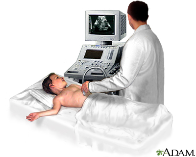
The test is done in the ultrasound or radiology department. A conducting paste is applied to your abdomen while you are lying down. The transducer (a hand-held instrument) is then moved over your abdomen.
There is little discomfort. The conducting gel may feel a little cold and wet. The sonographer may press the probe against your abdomen.
How you will prepare for the test depends on the problem. You will likely be asked not to eat or drink for several hours before the exam. Your health care provider will go over what you need to do.
You will be lying down for the procedure. A clear, water-based conducting gel is applied to the skin over the abdomen. This helps with the transmission of the sound waves. A handheld probe called a transducer is then moved over the abdomen.
Levine MS, Gore RM. Diagnostic imaging procedures in gastroenterology. In: Goldman L, Schafer AI, eds. Goldman-Cecil Medicine. 26th ed. Philadelphia, PA: Elsevier; 2020:chap 124.

You may need to change position so that the health care provider can look at different areas. You may also need to hold your breath for short periods during the exam.
Barnett CF, Sweeney DA, Huddleston LL. Ultrasonography: advanced applications and procedures. In: Broaddus VC, Ernst JD, King TE, et al, eds. Murray and Nadel's Textbook of Respiratory Medicine. 7th ed. Philadelphia, PA: Elsevier; 2022:chap 24.Chen L. Abdominal ultrasound imaging: anatomy, physics, instrumentation, and technique. In: Sahani DV, Samir AE, eds. Abdominal Imaging. 2nd ed. Philadelphia, PA: Elsevier; 2017:chap 3.Levine MS, Gore RM. Diagnostic imaging procedures in gastroenterology. In: Goldman L, Schafer AI, eds. Goldman-Cecil Medicine. 26th ed. Philadelphia, PA: Elsevier; 2020:chap 124.Nickels LC, Duran-Gehring P. Emergency ultrasound. In: Walls RM, ed. Rosen's Emergency Medicine: Concepts and Clinical Practice. 10th ed. Philadelphia, PA: Elsevier; 2023:chap e3.Wilson SR. The gastrointestinal tract. In: Rumack CM, Levine D, eds. Diagnostic Ultrasound. 5th ed. Philadelphia, PA: Elsevier; 2018:chap 8.
How you will prepare for the test depends on the problem. You will likely be asked not to eat or drink for several hours before the exam. Your health care provider will go over what you need to do.
Wilson SR. The gastrointestinal tract. In: Rumack CM, Levine D, eds. Diagnostic Ultrasound. 5th ed. Philadelphia, PA: Elsevier; 2018:chap 8.
You may have this test to: Find the cause of abdominal painFind the cause of kidney infectionsDiagnose or monitor tumors and cancersDiagnose or treat ascitesLearn why there is swelling of an abdominal organLook for damage after an injuryLook for stones in the gallbladder or kidneyLook for the cause of abnormal blood tests such as liver function tests or kidney testsLook for the cause of a fever The reason for the test will depend on your symptoms.
There is little discomfort. The conducting gel may feel a little cold and wet. The sonographer may press the probe against your abdomen.
The meaning of abnormal results depends on the organ being examined and the type of problem. Talk to your provider if you have any questions or concerns.An abdominal ultrasound can indicate conditions such as:Abdominal aortic aneurysmAbscessAppendicitisCholecystitisGallstonesHydronephrosisKidney stonesPancreatitis (inflammation in pancreas)Spleen enlargement (splenomegaly)Portal hypertensionLiver tumorsObstruction of bile ductsCirrhosis
Reviewed by: Jason Levy, MD, FSIR, Northside Radiology Associates, Atlanta, GA. Also reviewed by David C. Dugdale, MD, Medical Director, Brenda Conaway, Editorial Director, and the A.D.A.M. Editorial team.
Abdominal ultrasound is a type of imaging test. It is used to look at organs in the abdomen, including the liver, gallbladder, spleen, pancreas, and kidneys. The blood vessels that lead to some of these organs, such as the inferior vena cava and aorta, can also be examined with ultrasound.
An ultrasound machine makes images of organs and structures inside the body. The machine sends out high-frequency sound waves that reflect off body structures. A computer receives these waves and uses them to create a picture. Unlike with x-rays or CT scans, this test does not expose you to ionizing radiation.
The esophagus, stomach, large and small intestine, aided by the liver, gallbladder and pancreas convert the nutritive components of food into energy and break down the non-nutritive components into waste to be excreted.
The meaning of abnormal results depends on the organ being examined and the type of problem. Talk to your provider if you have any questions or concerns.
Yellow PagesTM, Walking Fingers & DesignTM, YP.caTM, YellowPages.caTM, Canada411TM, are trademarks of Yellow Pages Digital & Media Solutions Limited in Canada. All other trademarks are the property of their respective owners. Copyright © 2024 Yellow Pages Digital & Media Solutions Limited. All Rights Reserved. 6.95.0 (rev 20240910.1254)
Nickels LC, Duran-Gehring P. Emergency ultrasound. In: Walls RM, ed. Rosen's Emergency Medicine: Concepts and Clinical Practice. 10th ed. Philadelphia, PA: Elsevier; 2023:chap e3.
Abdominal ultrasound is a scanning technique used to image the interior of the abdomen. Like the X-ray, MRI, and CT scan, it has its place as a diagnostic tool. Ultrasound scans use high frequency sound waves to produce an image and do not expose the individual to radiation. The procedure is painless and safe.
Barnett CF, Sweeney DA, Huddleston LL. Ultrasonography: advanced applications and procedures. In: Broaddus VC, Ernst JD, King TE, et al, eds. Murray and Nadel's Textbook of Respiratory Medicine. 7th ed. Philadelphia, PA: Elsevier; 2022:chap 24.
This is the typical appearance of the blood vessels (vasculature) and urine flow pattern in the kidney. The blood vessels are shown in red and the urine flow pattern in yellow.
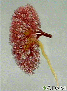
The kidneys are responsible for removing wastes from the body, regulating electrolyte balance and blood pressure, and the stimulation of red blood cell production.
Chen L. Abdominal ultrasound imaging: anatomy, physics, instrumentation, and technique. In: Sahani DV, Samir AE, eds. Abdominal Imaging. 2nd ed. Philadelphia, PA: Elsevier; 2017:chap 3.
An ultrasound machine makes images of organs and structures inside the body. The machine sends out high-frequency sound waves that reflect off body structures. A computer receives these waves and uses them to create a picture. Unlike with x-rays or CT scans, this test does not expose you to ionizing radiation. You will be lying down for the procedure. A clear, water-based conducting gel is applied to the skin over the abdomen. This helps with the transmission of the sound waves. A handheld probe called a transducer is then moved over the abdomen.You may need to change position so that the health care provider can look at different areas. You may also need to hold your breath for short periods during the exam.Most of the time, the test takes less than 30 minutes.


 Neil
Neil 
 Neil
Neil