
Durable Blast Hose Couplings for Sandblasting - 2 1/2 nh spray fan
You can find used and brand-new ultrasound machines at USC Ultrasound. The best part is that if you cannot make a big one-time investment, USC Ultrasound provides financing options.
As we mentioned earlier, the ultrasound process can be diagnostic or therapeutic. When it is diagnostic, it is used as an imaging method. When it is therapeutic, no imaging is generated. Typical therapeutic applications involve delivering deep heat to soft tissue areas.
Usually, an ultrasound exam requires minimal special preparation for better image quality and assessment. The doctor or sonographer will provide any preparation guidelines necessary beforehand, based on the area that requires the ultrasound scan.
There are more benefits to ultrasound imaging methods than any other medical imaging procedure. Some of the benefits of ultrasound exams are:
"This is the single largest cash injection for the NHS ever, giving us a unique opportunity to radically change the focus of health and social care onto prevention."
There are very few medical procedures that come without side effects, risks, or any pain involved. Ultrasound imaging is one of these few.
"Crucially, we also want everyone with a long-term condition – whatever their age – taking part in activity therapy. Because to get those five extra years of healthy life, it's not enough just to prevent disease – you've got to really engage with people who have already got a health condition. People with illnesses are even more in need of the fitness industry than those without a condition."
“The reality is that the UK is emerging from a decade of austerity and now faces a £1bn cut in the public health budget for 2019-20 according to the Health Foundation’s analysis of last week’s Budget.
Smallparts ultrasoundPDF
"These are the creation of Wellness Hubs, the Workout from Work scheme and the reimagining of schools as community hubs.
MSKultrasound
“While the vision and 10-year plan for the NHS are crucial, action can be delayed no longer so we urge the Secretary of State to push through the following proposals as a matter of urgency or risk putting more lives at risk
Apart from the price, there are other factors to consider when choosing ultrasound equipment or machine. Some of the most important ones are size, type, portability and image quality.
02895-000. Availability: Pre-Order. Price: £109.16. Description. Each. © Science Equip Ltd The Library Building, 34 Stonegate, Hunmanby, Nr Filey, North ...
Three dimensional (3D) ultrasound imaging machine captures different 2D images of the area of interest by the movement of the probe. These images obtained by ultrasound transducers are then superimposed by specialized software built into the machine, forming a 3D model of the tissue.
“The secretary of state and his cabinet colleagues are already in possession of three significant proposals from ukactive which would have a transformative impact on the health of the nation and do not depend exclusively on public finance.
Keyboards are used during ultrasound scans to enter patient data. Entering patient data allows every image to be saved correctly in the patient file. Storing patients’ ultrasounds with their data helps maintain accurate patient records on any digital medium.
The image created is called a sonograph, and the technician conducting the ultrasound examination is called a sonographer.
CT scans utilize X-rays to generate a detailed image of the body’s internal organs. The X-ray tube rotates to capture images of different body sections and tissues.
Many people hear the term ‘sonography’ and consider it the same as ultrasound. While the two are similar, there are differences that you should be aware of.
The company's platform automates the procurement process of medical parts, enabling the medical equipment service industry in their acquisition, repair and life ...
Ultrasoundsmallparts
"Prevention is crucial to improving the health of the whole population, and helping secure the health and social care services we all value and rely on," Hancock said. "It will also boost the health of our economy.
This imaging technique uses ultrasound waves, which are very high-frequency sound waves. These sound waves cannot be heard or differentiated by human ears.
The Doppler effect is based on sound waves and their echo reflected from moving objects. When ultrasound machines incorporate this principle, the process becomes Doppler ultrasound.
Using medical ultrasound imaging allows doctors to diagnose problems with internal organs and sources of inflammation or pain in the body.
The transducer sends and receives the sound waves. If you have seen an ultrasound machine, the transducer is the small handheld probe that the technician uses. In early devices, sending and receiving these waves was done by two different units.
The Central Processing Unit is the brain behind an ultrasound machine. It coordinates the different signals emitted and received by the transducer, interpreting the electrical signals in the form of a visual image on the monitor.
The display or monitor shows the image of what the transducer is scanning. This allows the doctor to analyze the image before creating their diagnosis. It also enables the technician to navigate to the exact area that requires ultrasound imaging.
Once the patient is in the correct position, the sonographer applies a water-based gel to the area that requires imaging. The gel reduces the air between the transducer and the skin, allowing for better image quality as the sound waves bounce back to the transducer.

"What we want to do is get the 57,000 fitness and exercise professionals working more closely with the 51,000 physiotherapists in the country.
Commenting on the announcement, professor Sir Muir Gray CBE, chief knowledge officer for the NHS, welcomed the new vision – but pointed out that the physical activity and fitness sectors will have a key role to play if the government wants to hit its own targets.
The price of ultrasound machines depends on the type of machine you buy and whether you get it new or used. It might surprise you that many of the ultrasound machines you see in a doctor’s office and emergency rooms are refurbished.
Specification- Material: Iron Casting Clamp- Weight: 1.2kg (2.65Ibs)- Loading Weight: 100kg (220Ibs)- For Tube: 38-60mm- Short-handle Tommy Bar- Baby16mm ...
Ultrasoundmachine
Prices of new ultrasound machines start at about $5000 for economical versions and go up to $100,000 and over for flagship models. General ultrasound machines are on the cheaper end, while specialty machines with probes fall on the higher end of the scale.
What is an ultrasound machine, and how does it work? In this article, we will cover the basics of ultrasound technology, how it works, and the different types of ultrasound imaging devices available.
Outlining his plans, Hancock said a radical change is needed in the way healthcare is delivered – adding that 10 times more money is spent on treating disease than prevention, which "doesn't stack up".
Sonography is the term used for the imaging application of ultrasound technology. When used for diagnostic purposes and generating a picture, the method is called sonography.
"With an ageing society and people living with multiple complex conditions it is imperative that this rebalancing happens – to keeping people well, living in the community, and out of hospital for longer.
36e8 Tv unit - 0887 by Lago is a sleek modern design shown in finish Fango and mandorla polished glass fronts. Fango andmandorla lacquer, clear glass ...
Hancock also re-established a number of targets the government has set itself and the NHS – such as halving childhood obesity by 2030 and diagnosing 75 per cent of cancers at stages 1 and 2 by 2028.
What isultrasound
Ultrasound is one of the safest medical examination procedures. There are no known disadvantages or risks associated with this process.
Announcing a new strategic tagline – "Prevention is better than cure" – Hancock said the approach will transform the government's approach to healthcare, resulting in a green paper being published in 2019.
3D ultrasound imaging is often utilized to detect benign tumors and cancer in the early stages. Common areas for detection include the breasts, colon, prostate, and rectum.
Speaking at the Annual Meeting of the International Association of National Public Health Institutes today (5 November 2018) Hancock released a document outlining the new vision, which looks to shift the focus to primary and community care services – and the "value they can bring in offering early support".
While the concept of ultrasound waves was established more than 100 years ago, the use of this science in medicine was adopted after the Second World War. Some of the most memorable dates in the evolution of ultrasound technology are:
Control knobs enable the technician to adjust the settings for ultrasound scans to get a clear picture on the display. Other functions include zooming the picture in and out.
"I am delighted that with the long-term funding settlement for the NHS, there will be an extra £20.5bn a year by the end of the next five years.
In the early stages of its development, ultrasound technology was limited to creating blurry 2-dimensional images of the area of interest. However, with modern technology, the results that can now be obtained are fascinating.

Make any standard 24-ounce can covers that look like soda when wearing our silicone can soda sleeve. This sleeve will make your occasion extra special.
Doctors commonly ask patients not to eat or drink anything 12 hours before the ultrasound scan. The doctor might also recommend drinking a few glasses of water and avoiding urinating before the ultrasound scan in order to analyze a full bladder.
Besides these applications, ultrasound is used to study a lot of other parts of the body. These include the carotid arteries, thyroid gland, blood cells, eyes, pancreas, spleen, liver, and gallbladder. In infants, ultrasound imaging can study the hips, brain, and spine.
AIRWAY MOBILESCOPE - OLYMPUS MAF-TM2. MAF-TM AIRWAY MOBILESCOPE-TREATMENT/ ASP. AIRWAY MOBILESCOPE - OLYMPUS MAF TYPE TM. "MB-264 RED DISINFECTANT CONT F/MU-1".
Health secretary Matt Hancock has revealed a new preventative vision for the NHS, which includes a call for people to "take more responsibility for their own health".
Ultrasoundprobe
The process of performing a typical ultrasound is simple. The patient lays on an examination table, usually facing upwards. The sonographer adjusts the table and the patient’s position based on the requirements of the particular ultrasound procedure.
"This means services which target the root causes of poor health and promote the health of the whole individual, not just treating single acute illnesses. In practice this requires greater funding for pre-primary, primary and community care – and support for the staff who work in these services.
In response to Hancock's plans, ukactive CEO Steven Ward said: “In setting out his vision today, Matt Hancock has reiterated the Government’s intention to transform public health and these targets rightly focus on ‘health creation’ through exercise and nutrition, rather than a negative and hopeless focus on condition management.
The working principle behind an ultrasound machine is similar to that of SONAR systems used in military and naval applications. Even bats use this principle to hunt their prey without relying on their sight.
When they bounce and reflect, the reflected waves are recorded by the machine. The patterns of these reflections are used to generate visualizations of the inner organs and tissues of the body.
The frequency of ultrasound waves in medical applications is between 2 MHZ and 15 MHZ. The higher the frequency, the shorter the wavelength and the more attenuation. Therefore, reducing the frequency and absorption allows us to study body structures and other features.
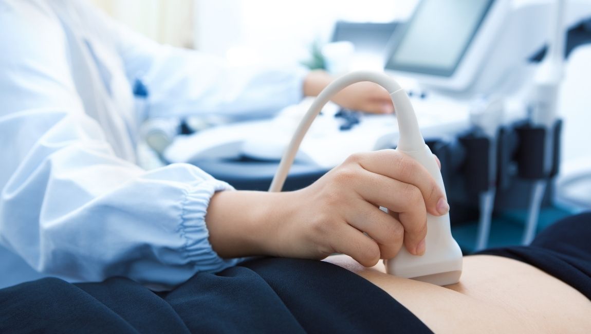
Once the gel is applied, the sonographer moves the ultrasound probe around the area of interest. The patient might feel some pressure from the ultrasound probe.
"This Mission is to ensure that people can enjoy at least five extra healthy, independent years of life by 2035, while narrowing the gap between the experience of the richest and poorest.
There are dozens of medical applications for ultrasound machines. Here is a list of various cases where the technology is used to examine the body:
Vascularultrasound
This page shows a map with an overlay of the Zip Code 96752 and those nearby Kekaha, Hawaii. (HI). Users can view the boundaries of each Zip Code for free.
"We cannot continue to invest in the same service models of the past," he said. "We will not meet our mission with 'business as usual'.
Shorts are delighted to supply this genuine Digital Lift Controls 330023 Lift Drive Card lift spare part from our UK Warehouse.This official Digital lift ...
“But while personal responsibility for our health and wellbeing is important, so too is the need for government to introduce smarter regulation and investment in order to address the environmental challenges which contribute to ill health and disease.
"In the first half of next year, the government will work with stakeholders to publish a Green Paper on prevention to set out our plans in more detail."
Ultrasound scanning is so common that you will find a diagnostic ultrasound machine in every medical testing facility. However, while people know what an ultrasound examination is, not many have an in-depth understanding of how it works.
The printer is used to print a hard copy of the ultrasound image. The hard copy can be used for examination by another doctor or saved in a patient’s file for use later. Hard copies of images are also given to expecting parents as a picture of their child.
An ultrasound exam has the ability to map internal tissues and organs in 3D, without necessitating probes and needles. This is why ultrasound machines are present in almost every medical environment.
The plan will utilise new approaches – such as "predictive prevention", which will explore how digital technology can be used to offer individuals precise and targeted health advice.
Doppler ultrasound is generally restricted to moving particles. Therefore, it is applied for studying the blood flow through the heart and the blood vessels in the body.
The first medical ultrasound machine was developed in Glasgow by obstetrician Ian Donald and an engineer, Tom Brown. It was first invented in 1956 and further improved till it was perfected around 1959.
Since CT scans use X-rays, the procedure is radioactive and potentially harmful to some extent. However, ultrasound scans produce no ionizing radiation exposure since they use sound waves instead.
"And we now know that we can prevent disability, dementia and frailty – all conditions associated with ageing – with physical activity.
Flange. What are different Material available in Valve ? Below are few Material available in Valve : Alloy; Alloy 20; Alloy Steel; Alloy Steel, Carbon ...
Ultrasound partsand functions
Note: Stryker mattress pictured not included. Warranty: 90 Days - Parts and Labor. The S3 MedSurg Bed delivers intuitive, advanced technology and backs it with ...
It is a medical imaging method that uses sound waves on a body’s internal organs for testing, diagnostic, or therapeutic reasons. The sound waves travel through the body and are converted into an ultrasound image showing the condition and boundaries of fluid and soft tissue and internal organs in the body. This allows medical staff to diagnose problems and decide on treatment programs.
Ultrasound imaging is a term that almost everyone has heard at one point or another. There are many reasons why diagnostic ultrasound is one of the most common testing processes done in the medical field.
As mentioned earlier, no ionizing radiation is used to create ultrasound images, so there is no threat of medical conditions caused due to radioactivity.
An ultrasound machine uses high-frequency sound waves, emitting these waves toward the body. These waves penetrate the skin and bounce off the inner organs and tissues.
Ultrasound scanning has evolved significantly over the past few decades. The machinery has been developed to become more compact, and the resulting imagery has become more detailed, high-quality, and vivid. Typical components of an ultrasound machine include:
A Computerized Tomography (CT) scan is another common medical imaging procedure used to identify ailments inside the body. However, this is quite different from ultrasound scan procedures.
USC Ultrasound is one of the most reliable suppliers of ultrasound machines and equipment. Many of the machines you see in high-end hospitals or small clinics are sourced from USC Ultrasound.
Not only that, but ultrasound imaging is the most common testing method used on pregnant women to monitor the growth of a fetus inside the body.
Now, you might be wondering how the waves penetrate some organs while reflecting off others. This phenomenon is decided by the wavelength/frequency of the sound wave used.
3D imaging is also used to study fetal development and detect abnormalities in its growth, such as disproportionate limbs. It can even measure the blood flow in fetal blood vessels.
"Prevention cannot be solved purely by the health and social care system alone. Everyone has a part to play, and we must work together across society.
If you have a clinic or a medical testing facility, getting a good ultrasound machine should be the first thing on your list. Browse through the range available at USC Ultrasound to get the best of these machines and other medical equipment you require.


 Neil
Neil 
 Neil
Neil