
PREMIERE Ice Melt Compounds & Traction Control - premiere ice melter
The meaning of GATCH BED is a hospital bed with a frame in three movable sections equipped with mechanical spring parts that permit raising the head end, ...
Cardiac Imaging 1 introduces these questions (and answers!), and much more. Develop an understanding of how emergency medicine and cardiology applications differ, and test your knowledge of different views of the heart.
The following point-of-care ultrasound courses are recommended for you based on what we know about your interests. You can follow the suggested curriculum, or explore other materials at your own pace. To view other available courses, click the courses button at the top of this page.
Determination of Stroke Volume (SV) is perhaps the most essential of all the “advanced” techniques for point-of-care echocardiography. It’s considered advanced, because it relies on quantitative spectral Doppler techniques, which are not routinely part of the core point-of-care echocardiography curricula offered for most specialties at the time of this publication. This technique is the most practical and intuitive gateway to hemodynamic understanding of echocardiography and is a powerful adjunct in the assessment of Left Ventricular (LV) function.
Gain an understanding of the procedures and techniques used to place a peripheral line using ultrasound guidance. Learn to identify adjacent anatomical structures and how to master the correct equipment settings for the procedure.
There are a number of different data points that can be collected that inform us of the function and loading conditions of the Right Ventricle (RV): Shape and size of the RV, Inferior Vena Cava (IVC), Tricuspid Angular Plane Systolic Excursion (TAPSE), and Right Ventricular Systolic Pressure (RVSP).
The axillary block is a plexus block at the terminal branches of the brachial plexus, designed to anesthetize the major motor and sensory nerves in the distal arm, elbow, wrist, forearm, hand, and fingers. Course participants will learn the anatomy of the axillary vessels and musculocutaneous nerve and the technique for performing an axillary nerve block.
Número de producto 1630MD · Descripción Llave de control, empaque comercial en caja · Cantidad por caja 50 · Ancho del cuerpo 1-7/8 in (48 mm) · Color Black.
What is an Evacuated Container used for
After completing the aorta ultrasound course, participants should be able to: identify the anatomical structures visualized during the aorta examination, recognize the various types of abdominal aortic aneurysms and perform their associated measurements, and determine the preferred transducer to perform the aorta exam.
NEET SS 2021 | DM Paediatrics - Rank 100, Dr Meghna Nema from SPEED #SPEEDSS #SpeedLearningApp #NEETSS Android: ...
Consolidation of infusion data into the patient electronic record was not feasible at the time with any vendor, therefore, the CareFusion Alaris met the ...
This course will compare Transthoracic Echocardiography (TTE) and Transesophageal Echocardiography (TEE), outline clinical questions with point-of-care TEE, describe how to get started with TEE (politics, cost, logistics), discuss safety and training, review a suggested TEE protocol, and give several case examples using TEE.
This introduction to Doppler principles and how they relate to point-of-care echocardiography will frame the knowledge you need to engage some particularly valuable hemodynamic techniques!
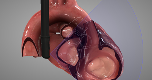
Point of care Transesophageal Ultrasound (TEU) is an emerging modality that offers bedside clinicians the ability to assess critically ill patients quickly. A Transesophageal Echocardiogram, or TEE, is an alternative way to perform an echocardiogram. A specialized transducer containing an ultrasound transducer at its tip is passed into the patient's esophagus. This allows image and Doppler evaluation which can be recorded.
Baxter
Order Tripp Lite SMART1000LCD (TL360-ND) at DigiKey. Check stock and pricing, view product specifications, and order online.
AFCO Distribution · LIVESTOCK · Livestock Feed · Other; GH SOYBEAN MEAL 46.5% 50#. Customer Login. Forgot My Password. Account Details. Welcome.
8x2 Treated Timber [Various Lengths] from 15.00INC VAT. Showing 1 of 1 Items. Be The First To Know Sign up for the Next newsletter today.
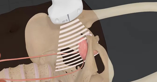
How do you angle the transducer to get a Parasternal Long Axis (PLAX) view of the heart in the Emergency Department? Which kind of transducer should you use? How is the cardiology orientation different? And how, exactly, do you measure fluid responsiveness using the Inferior Vena Cava (IVC)?
Doppler echocardiography is the language of flow in and around the heart. In order to evaluate hemodynamics in and around cardiac valves, cardiac pressures or in the calculation of Stroke Volume (SV), one must speak the language of Doppler.
FUJIFILM Sonosite, Inc. The Sonosite logo and other trademarks not owned by third parties are registered and unregistered trademarks of FUJIFILM Sonosite, Inc. in all jurisdictions. All other trademarks are the property of their respective owners.
Evacuated container meaning
Welcome to PFR, We are your one stop shop to finding those F1 Parts and F1 Memorabilia you thought you would never find, let alone own!
6 days ago — ... GE Healthcare to procure medical equipment Agencies. Representative Image. Synopsis. KIMS has signed an MoU with Wipro GE Healthcare to enhance ...
8888266197 Anti-Reflux Valve, 10/cs 60210 CB-X I Cleaning Brush, Single Ended, 5.0mm, 50/cs By removing the transitional connector, the same sets will 60218 ...
Central Line placement is one of the most common hospital procedures, and yet it is not without risk. Ultrasound-guided Central Venous Catheter (CVC) placement reduces some of the more severe risks, such as pneumothoraces and CLABSI. Learn the procedures, techniques, and best practices associated with placing a central line under ultrasound guidance while minimizing risks of iatrogenic complications.


 Neil
Neil 
 Neil
Neil