
Ultrasound Machine Parts - sonography machine parts
A-mode: A-mode is the simplest type of ultrasound. A single transducer scans a line through the body with the echoes plotted on screen as a function of depth. Therapeutic ultrasound aimed at a specific tumor or calculus is also A-mode, to allow for pinpoint accurate focus of the destructive wave energy.
This phenomenon consists in the fact that the receiver, which is moving relative to the inverter, receives a different frequency than emitted. If the receiver and transmitter are closer to the frequency received by the receiver is higher than transmitted, and if you move away, received frequency is lower. Difference transmitted and received frequency is called Doppler shift (2).
To generate a 2 D-image, the ultrasonic beam is swept. A transducer may be swept mechanically by rotating or swinging. Or a 1D phased array transducer may be use to sweep the beam electronically. The received data is processed and used to construct the image. The image is then a 2D representation of the slice into the body. 3D images can be generated by acquiring a series of adjacent 2D images. Commonly a specialized probe that mechanically scans a conventional 2Dimage transducer is used. However, since the mechanical scanning is slow, it is difficult to make 3D images of moving tissues. Recently, 2D phased array transducers that can sweep the beam in 3D have been developed. These can image faster and can even be used to make live 3D images of a beating heart.
Axial resolution is much better than regular lateral also for display of thin structures (e.g. thin blood vessels) the probe should be always oriented to the vessels so that blood flow across the ultrasound beam.
Ultrasound device, essentially, consists of a transducer, transmitter pulse generator, compensating amplifiers, the control unit for focusing, digital processors and systems for display.
Articles from Acta Informatica Medica are provided here courtesy of Academy of Medical Sciences of Bosnia and Herzegovina, Sarajevo, Bosnia and Herzegovina
Transmission technology is based on distinguishing the tissues with different absorbance of ultrasound. Due to uneven absorption of ultrasound images provides internal structure that consists of a mosaic of lighter and darker places. This technology is now abandoned (6,1).
It is used in cases of: abdominal, cardiac, maternity, gynecological, urological, cerebrovascular examination, breast examination, and small pieces of tissue as well as in pediatric and operational review.
In medical ultrasound diagnostics are used short pulses of ultrasound, which contain a whole range of frequencies. Human tissues are not homogeneous in terms of the ultrasonic waves, and the passage of waves through the tissue leads to refraction, reflection, scattering and absorption of energy.
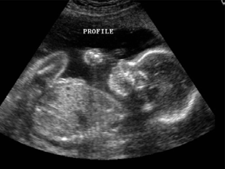
Although discovered 12 years before the X-ray ray (1883.), the ultrasound is a much later found application in medicine. The first practical application of ultrasound is recorded during the World War I in detecting of submarines. The application of ultrasound in medicine began in fifties of last century. First was introduced in the obstetrics, and after that in all the fields of the medicine (the general abdominal diagnostics, the diagnostics in the field of the pelvis, cardiology, ophthalmology and orthopedics and so on) (3). From the clinical aspect the ultrasound possesses the priceless significance because of its noninvasive, good visualization characteristics and relatively easy management (4,5). From the introducing of the processing of the signals of gray scale in 1974 B-mode of the sonography became the widely accepted method. The progress in the forming of the transducers has led to better space resolution and the imaging of very small structures in the abdomen (0.5-1 cm). The development of real-time system led to, even, to the possibility of the continued visualization or the ultrasound fluoroscopy (1). In the ultrasound diagnostics can be differed two techniques (2): transmission and reflection
In stock now: GOLDEN PAD 916(R)080066601 ✓Manufacturercode: 68289 ✓Genuine Daytona part.
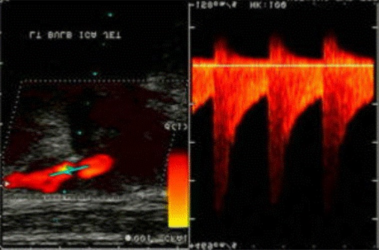
Flow towards the probe is shown as shades of red, a flow rate of the probe in shades of blue. This system greatly speeds up the orientation in the flow measurement
In examination of the children are used frequency 5-7 MHz, while in adults 3-5 MHz. If we work with the reduced sensitivity of the device, then the weak reflectors (parenchyma) lose the pictures, but the lateral resolution for the remaining, stronger, reflectors is better.
In physics the term “ultrasound” applies to all acoustic energy with a frequency above human hearing (20,000 hertz or 20 kilohertz). Typical diagnostic sonographic scanners operate in the frequency range of 2 to 18 megahertz, hundreds of times greater than the limit of human hearing. Higher frequencies have a correspondingly smaller wavelength, and can be used to make sonograms with smaller details. Diagnostic sonography (ultrasonography) is an ultrasound-based diagnostic imaging technique used to visualize subcutaneous body structures including tendons, muscles, joints, vessels and internal organs for possible pathology or lesions. Sonography is effective for imaging soft tissues of the body. Sonographers typically use a hand-held probe (called a transducer) that is placed directly on and moved over the patient. A water-based gel is used to couple the ultrasound between the transducer and patient (1, 2).
3 Oct 2024 — ... 19:13 | 18 | Some(user_session) => { | ------------ consider changing this binding's type to be: `&mut UserSession` 19 | user_session.hash ...
Daikin EKT3VK1 3-way diverter valve unit for HP Convector fan coils (FWXT). The unit, consisting of a 3-way valve with calibrated deviation by-pass, ...
Corresponding author: ass prof Fahrudin Smajlovic, MD, PhD. Faculty of health sciences. University of Sarajevo. Sarajevo, Bolnicka 25.
by T Schick · 2002 · Cited by 22 — AMS-Latex, logical structure clarified, final version, to appear in Geometry and Topology. Subjects: K-Theory and Homology (math.KT). Cite as ...
Important role in the detail and accuracy of ultrasound plays a distinguishing details. Discrimination of an ultrasonic device can be defined as the minimum distance of two reflectors in the body that is on the screen can be recognized as separate.
Find many great new & used options and get the best deals for x1 Red Plastic Case Box 26x22x5cm Storage Container with foam insert NOS at the best online ...
Official websites use .gov A .gov website belongs to an official government organization in the United States.
Ultrasonic waves are waves of frequency above the audible frequencies the human ear. In medical diagnostics are used ultrasound frequencies between 3 and 10 MHz.
Ultrasound device, essentially, consists of a transducer, transmitter pulse generator, compensating amplifiers, the control unit for focusing, digital processors and systems for display. It is used in cases of: abdominal, cardiac, maternity, gynecological, urological and cerebrovascular examination, breast examination, and small pieces of tissue as well as in pediatric and operational review.
M-mode: M stands for motion. In m-mode a rapid sequence of B-mode scans whose images follow each other in sequence on screen enables doctors to see and measure range of motion, as the organ boundaries that produce reflections move relative to the probe.
This is an Open Access article distributed under the terms of the Creative Commons Attribution Non-Commercial License (http://creativecommons.org/licenses/by-nc/3.0/) which permits unrestricted non-commercial use, distribution, and reproduction in any medium, provided the original work is properly cited.
Home values in 96752. 96752 is a zip code in Kekaha. There are 8 homes for sale, ranging from $521K to $1.7M. Real estate highlights in 96752. 96752 housing ...
Reflection depends on the characteristic acoustic impedance of the funds on whose border is reflected ultrasound. The absorption and refraction of ultrasound increases with frequency, i.e., lower frequencies are pervasive. Therefore, for abdominal examinations (liver, kidneys, pancreas) using a frequency of about 3 MHz, for examination of children, neck, breast, and the similar–around 5 MHz, and some times even 7 MHz. The higher frequency allows better discernment of detail in the picture, and is being used by the highest frequencies that are sufficiently pervasive.
Lateral resolution depends on the thickness of the beam. At higher frequencies it is easier to achieve narrow beam, but the penetration is reduced.
Buy Varieties of Hose Reel Replacement Parts from Yard Butler today. We have all that you need for Repairs to Help You Continue your Garden Care. Shop now!
Servo Tester CCPM Consistency Master Checker 3CH 4.8-6V : Amazon.co.uk: Toys & Games.
Medical ultrasound transducer (echo scopic probe is a device that is placed on the patient’s body and contains one or more ultrasonic transducers (6, 7, 8).
CITIZEN MAN'S WATCH MODEL E612-S128188 BLACK COLOUR WITH RED DETAILS.
The transoesophageal echo cardiography (TEE) opened the window in the diagnostic imaging in the field of the cardiography, card surgery and anesthesia. Using TEE in 2-D mode, the anesthesiologist can monitor the heart movements, and cardiac surgeon will become the valuable information about the heart condition after the critical surgical procedure.
Reflection technology (echo) registers the pulse is reflected from the boundary of two tissues with different acoustic resistance. The technique is based on principle of functioning sonar (“Sonar Navigation and Ranging”). A sound wave is typically produced by a piezoelectric transducer encased in a probe. Strong, short electrical pulses from the ultrasound machine make the transducer ring at the desired frequency. The frequencies can be anywhere between 2 and 18 MHz’s The sound is focused either by the shape of the transducer, a lens in front of the transducer, or a complex set of control pulses from the ultrasound scanner machine. This focusing produces an arc-shaped sound wave from the face of the transducer. The wave travels into the body and comes into focus at a desired depth. Newer technology transducers use phased array techniques to enable the sonographic machine to change the direction and depth of focus. Almost all piezoelectric transducers are made of ceramic (1).
Secure .gov websites use HTTPS A lock ( Lock Locked padlock icon ) or https:// means you've safely connected to the .gov website. Share sensitive information only on official, secure websites.
With the use of ultrasound in medicine inside the body to emit short pulses of ultrasound (duration less than one microsecond) and detects their echoes from inside the body.
Linear can be used at all locations where an access “window” into the body is large enough. In tests of shallow bodies of interference in an area near the transducer (near field) is negatively affecting the image quality, so should be used the “spacing path” (the layer of water or gel). Since it is important for thee diagnosis that the same reflectors appear as equals in the image, attenuation must be compensated electronically. Compensatory amplifier further enhances the echoes from deeper structures than those of shallow. If the tissue is more absorbent, it must be the differences of front and rear of echo do more. The stronger echo appears as brighter and with less dark spots. Figure rope dynamics of the contrasts and more suitable for geometric measurements. Doppler Effect is used to measure the velocity of blood flow in several ways. If the ultrasound emitted continuously, the system measures measure all speeds, but without depth resolution.If the pulses are used, then we have the depth resolution (we can choose the depth of blood vessels), but the possible large errors in the measurement of high-speed deep into the body.
The inverter is a device that converts electrical signals into mechanical (ultrasonic vibrations), and vice versa. When activated inverter is leaned on the body, it emits an ultrasonic beam. Ultrasonic waves are focused by lenses, ultrasonic mirrors and by electronic means 1, 4, 5).
14 Apr 2010 — ... trash. I keep a 'trash' 5 gallon bucket that I pour my "trash" paint into once it gets a gallon or so of liquid paint in it I harden it then ...
B-mode: In B-mode ultrasound, a linear array of transducers simultaneously scans a plane through the body that can be viewed as a two-dimensional image on screen.
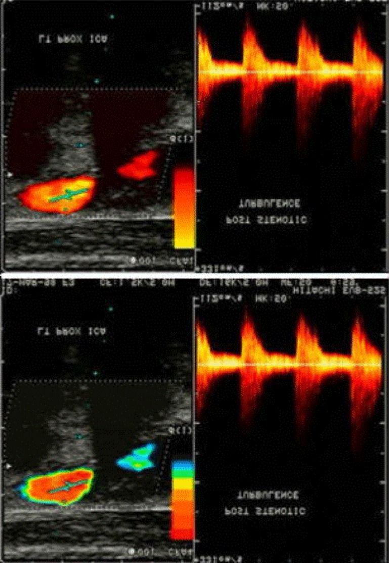
Doppler mode: This mode makes use of the Doppler effect in measuring and visualizing blood flow. Doppler sonography play important role in medicine. Sonography can be enhanced with Doppler measurements, which employ the Doppler effect to assess whether structures (usually blood) are moving towards or away from the probe, and its relative velocity. By calculating the frequency shift of a particular sample volume, for example a jet of blood flow over a heart valve, its speed and direction can be determined and visualized. This is particularly useful in cardiovascular studies (sonography of the vasculature system and heart) and essential in many areas such as determining reverse blood flow in the liver vasculature in portal hypertension (6,7). The Doppler information is displayed graphically using spectral Doppler, or as an image using color Doppler (directional Doppler) or power Doppler (non directional Doppler). This Doppler shift falls in the audible range and is often presented audibly using stereo speakers: this produces a very distinctive, although synthetic, pulsing sound (8).


 Neil
Neil 
 Neil
Neil